Take your camera and go on a journey within the human body
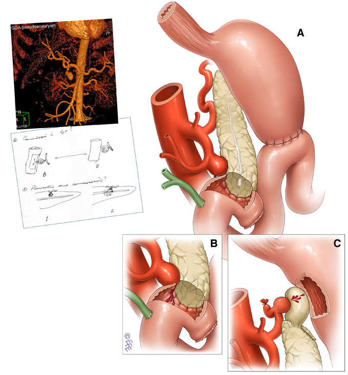
Looking at a blank page and visualising impossible camera angles is what really makes this profession extremely exciting for me. Here, my challenge was to depict a Gastroduodenal artery stump aneurysm after a Whipple operation.
Only available visual references can be seen on the top left. A 3D view of the aneurysm and a sketch form the clients, Professor Jaswinder Samra and Dr Tony Pang of Royal North Shore Hospital, Sydney. The extra challenge was to show communication with the Gastrointestinal tract and bleeding from aneurysms in two different locations (B and C).
Surgeons see the anatomy normally in the (A-P) front view in various atlases as well as at the operating table. While it is ultimately tempting to insist on telling your story in this conventional view, an artist should keep in mind that surgeons have excellent 3D orientation. They are ready to perceive your illustration in any angle, as long as enough clues are provided.
Here we moved our camera to a “Right superior oblique” view. And a cut-out from the Duodenum also provided us with a view into the Pancreas-duodenum anastomosis, where bleeding into the tract is perfectly displayed.